A CT scan is one of the fastest and most informative tests a doctor can order. In a few minutes, it produces detailed cross-sectional pictures of the bones, organs, blood vessels and soft tissues inside your body, helping doctors find problems that a regular X-ray may miss.
Doctors recommend a CT scan when they need a clear, quick answer: after an accident, when someone has sudden severe abdominal pain, a persistent cough, unexplained weight loss, or a sudden severe headache. It is also used to plan surgery, guide biopsies and check whether a treatment is working.
The three most commonly requested scans are the chest CT scan, the CT scan abdomen and the CT brain scan. Each looks at a different part of the body and answers different clinical questions.
In this guide, you will learn what a CT scan is and how it works, the main types and their uses, how to prepare, what happens on the day, the difference between scans with and without contrast, how safe CT scans are, how CT compares with X-ray and MRI, and when you can expect your report.
What Is a CT Scan?
CT stands for computed tomography. It is also called a CAT scan (computed axial tomography). A CT scan uses X-rays and a computer to create detailed cross-sectional images, or "slices", of the body.
How does a CT scanner work?
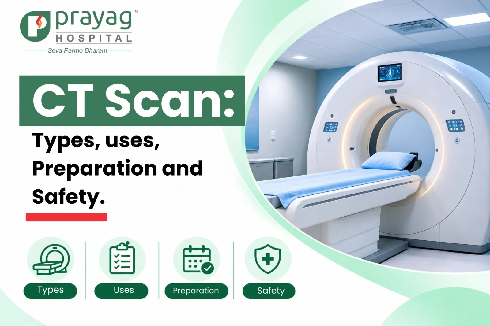
A CT scanner is a large, ring-shaped machine with a short tunnel in the middle. You lie on a motorised table that slides slowly through the ring. Inside, an X-ray tube rotates around you while detectors on the opposite side measure how much X-ray energy passes through each part of the body.
A computer turns these measurements into thin slices. Think of it like slicing a loaf of bread and looking at each slice separately. The slices can also be stacked to create 3D images, which help doctors see the exact size, shape and location of a problem.
What can a CT scan help detect?
A CT scan can help doctors identify:
Fractures & Injuries
Injuries to bones, joints and internal organs.
Internal Bleeding
Bleeding inside the skull, chest or abdomen.
Tumours
Tumours and the spread of cancer.
Lung Conditions
Infections such as pneumonia, and lung diseases such as emphysema.
Blood Clots
Including clots in the lungs (pulmonary embolism).
Kidney Stones
Kidney stones and urinary tract blockages.
Abdominal Emergencies
Appendicitis, diverticulitis and bowel obstruction.
Brain Conditions
Stroke and other brain conditions.
CT is also used to guide biopsies and drainage procedures, plan surgery and radiotherapy, and monitor how a disease responds to treatment.
CT scan vs regular X-ray
A regular X-ray takes a single flat (2D) picture, in which organs overlap one another. A CT scan takes hundreds of images from many angles and builds detailed slices, so structures do not hide behind each other. This makes CT far better at showing soft tissues, blood vessels and small abnormalities. The trade-off is that a CT scan uses a higher radiation dose than a single X-ray, which is why doctors order it only when the extra detail is needed.
Types of CT Scans
CT scans are named after the part of the body being examined. Your doctor chooses the type based on your symptoms and the question they need answered. The three most common are explained below.
Chest CT Scan
What is a chest CT scan? A chest CT scan takes detailed images of the lungs, heart, major blood vessels, airways, ribs and the lymph nodes in the chest.
Why do doctors recommend it? It is advised for a long-standing cough, coughing up blood, breathlessness, chest pain, an abnormal chest X-ray, or to follow up a known lung condition.
Conditions it can help identify:
- Pneumonia, tuberculosis and other lung infections
- Lung nodules and lung cancer
- Pulmonary embolism (a blood clot in the lungs)
- Interstitial lung disease, emphysema and bronchiectasis
- Fluid around the lungs and chest injuries
Chest CT scan vs chest X-ray: A chest X-ray is quick, low-dose and often the first test. A chest CT shows far more detail and can pick up small nodules, early infections or clots that an X-ray may miss. A standard chest CT gives about 7 mSv of radiation, compared with about 0.1 mSv for a chest X-ray, so it is used when the extra detail will change your care. A low-dose chest CT, used for lung screening, gives about 1.5 mSv.
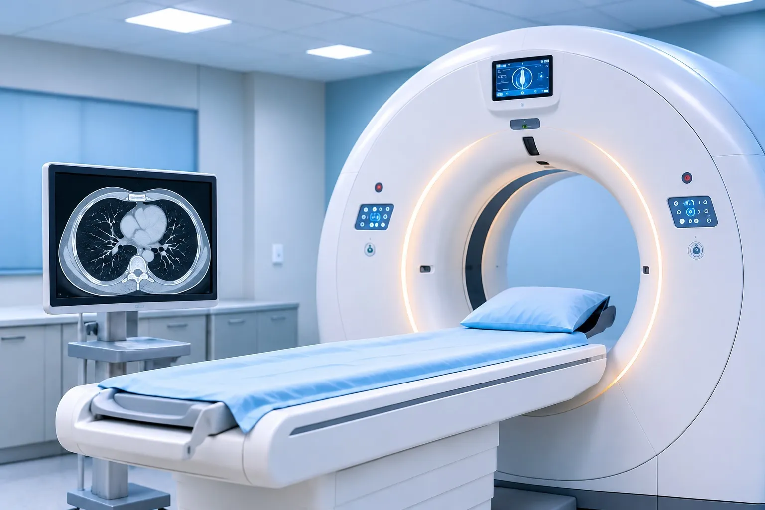
How to prepare: Most plain chest CT scans need no fasting. If contrast is planned (for example, to check for a clot), you may be asked not to eat for a few hours. You will be asked to hold your breath for a few seconds during the scan.
CT Scan Abdomen
What is an abdominal CT scan? A CT scan abdomen examines the organs in the belly. It is often combined with the pelvis and called a CT abdomen and pelvis.
Organs it can examine: liver, gallbladder, pancreas, spleen, kidneys, adrenal glands, stomach, small and large intestine, appendix, urinary bladder, reproductive organs and major blood vessels.
Conditions it can help detect:
- Appendicitis, diverticulitis and bowel obstruction
- Kidney stones and urinary blockages
- Pancreatitis and gallbladder problems
- Liver lesions, abscesses and tumours
- Internal bleeding or organ injury after an accident
- Hernias and enlarged lymph nodes
When is contrast required? Contrast is often used to assess tumours, infections, abscesses, inflammation, blood vessels and injuries. It may be given through a vein, as a drink, or both. A plain (non-contrast) CT is usually enough for kidney stones.
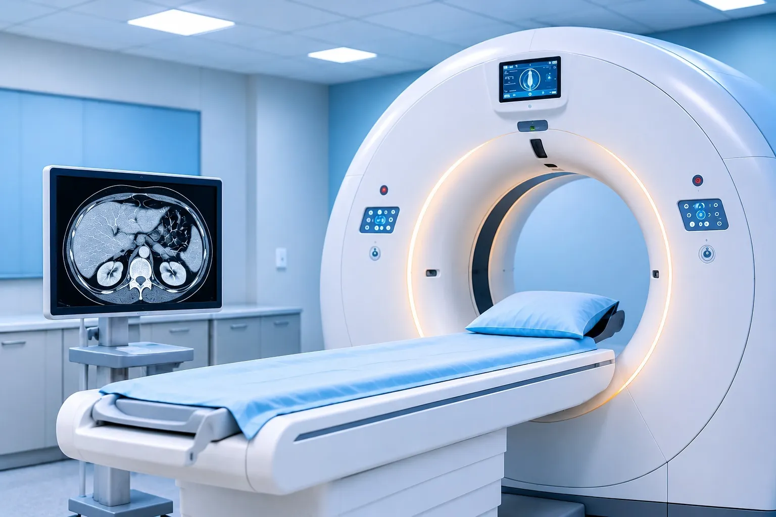
How to prepare: You may be asked to fast for 4 to 6 hours. If oral contrast is needed, you will drink it over about an hour before the scan, so arrive early. Bring your recent kidney function test (serum creatinine) if contrast is planned.
CT Brain Scan
What is a CT brain scan? A CT brain scan (also called a CT head scan) creates images of the brain, skull, sinuses and surrounding structures.
Why do doctors recommend it? It is commonly advised for a head injury, a sudden severe headache, suspected stroke, seizures, sudden confusion, fainting with injury, or sudden weakness, numbness or loss of speech.
Head injury and suspected bleeding: CT is the first-choice test to check for bleeding inside the skull after a head injury, because it is quick and very good at showing fresh blood and skull fractures.
- Has lost consciousness or cannot remember events around the injury
- Vomits more than once
- Has a seizure, weakness, or blurred or double vision
- Has signs of a skull fracture, such as clear fluid from the nose or ears
- Is elderly, or takes blood thinners
CT brain scan vs MRI: A CT brain scan takes a few minutes and is ideal in emergencies. MRI takes longer (often 30 to 45 minutes) and uses no radiation. It shows finer detail of brain tissue, so it is preferred for small tumours, multiple sclerosis, epilepsy work-up and early stroke changes. Your doctor may order both.
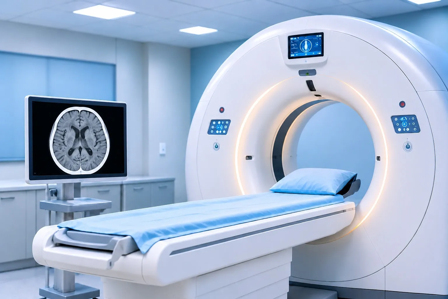
How to prepare: A plain CT brain scan needs no special preparation. Remove hairpins, earrings, spectacles and dentures if asked. Fasting is needed only if contrast is planned.
How to Prepare for a CT Scan
Good preparation makes the scan safer and the images clearer. Your hospital will give you specific instructions, but these general steps apply to most patients.
Is fasting required? For a plain CT scan, usually not. For a scan with contrast, especially a CT scan abdomen, you may be asked to avoid food for 4 to 6 hours. Small sips of water are generally allowed. Continue your regular medicines unless your doctor advises otherwise.
What should you wear? Loose, comfortable clothes without metal zips, buttons or hooks. Leave jewellery at home. You may be asked to change into a hospital gown and remove hairpins, spectacles, hearing aids and removable dentures.
What Medical Records Should You Bring?
- Your doctor's prescription or referral for the CT scan
- Previous CT, MRI, X-ray or ultrasound reports and images
- A recent kidney function test (serum creatinine) if contrast is planned
- A list of current medicines
- A photo ID and any insurance or TPA documents
What Should You Tell the Doctor Before the Scan?
- If you are, or might be, pregnant, or if you are breastfeeding
- Any allergy, especially a previous reaction to contrast dye
- Kidney disease, diabetes, asthma or thyroid problems
- If you take metformin for diabetes, or blood thinners
- If you have a fear of closed spaces
Precautions for contrast: Your team will check your kidney function and allergy history before injecting contrast. Patients with a past mild reaction may be given medicines beforehand. Drink plenty of water after the scan, unless you have been told to limit fluids.
What Happens During a CT Scan?
Registration and Checks
You arrive, complete a short form and a technologist confirms your details, allergies and pregnancy status.
Changing
You remove metal objects and may change into a gown.
IV Line (If Needed)
For a contrast scan, a small cannula is placed in a vein in your arm.
Positioning
You lie on the scanner table, usually on your back. Pillows or straps help you stay still.
Scanning
The table moves slowly through the ring. You may hear a soft whirring sound. The technologist watches from the next room and speaks to you through an intercom, asking you to hold your breath for a few seconds.
Contrast Injection
If used, you may feel a warm, flushing sensation or a metallic taste for a few seconds. This is normal.
Finish
The table moves out and the cannula is removed.
Is a CT scan painful? No. The scan itself is painless. The only discomfort may be the needle prick for contrast or lying still on a firm table.
How long does a CT scan take? The actual scanning usually takes less than five minutes. Including preparation, the full appointment can take 15 to 30 minutes, and up to an hour or more if oral contrast is needed.
What happens after the scan? Most people go home and return to normal activities straight away. If you had contrast, you may be asked to wait for a short time to make sure you feel well, and to drink extra water over the next day to help flush it out.
CT Scan With Contrast vs Without Contrast
What is contrast material? Contrast (often called dye) is a special liquid that shows up brightly on CT images. For CT, it is usually iodine-based and given through a vein. Some abdominal scans also use contrast you drink.
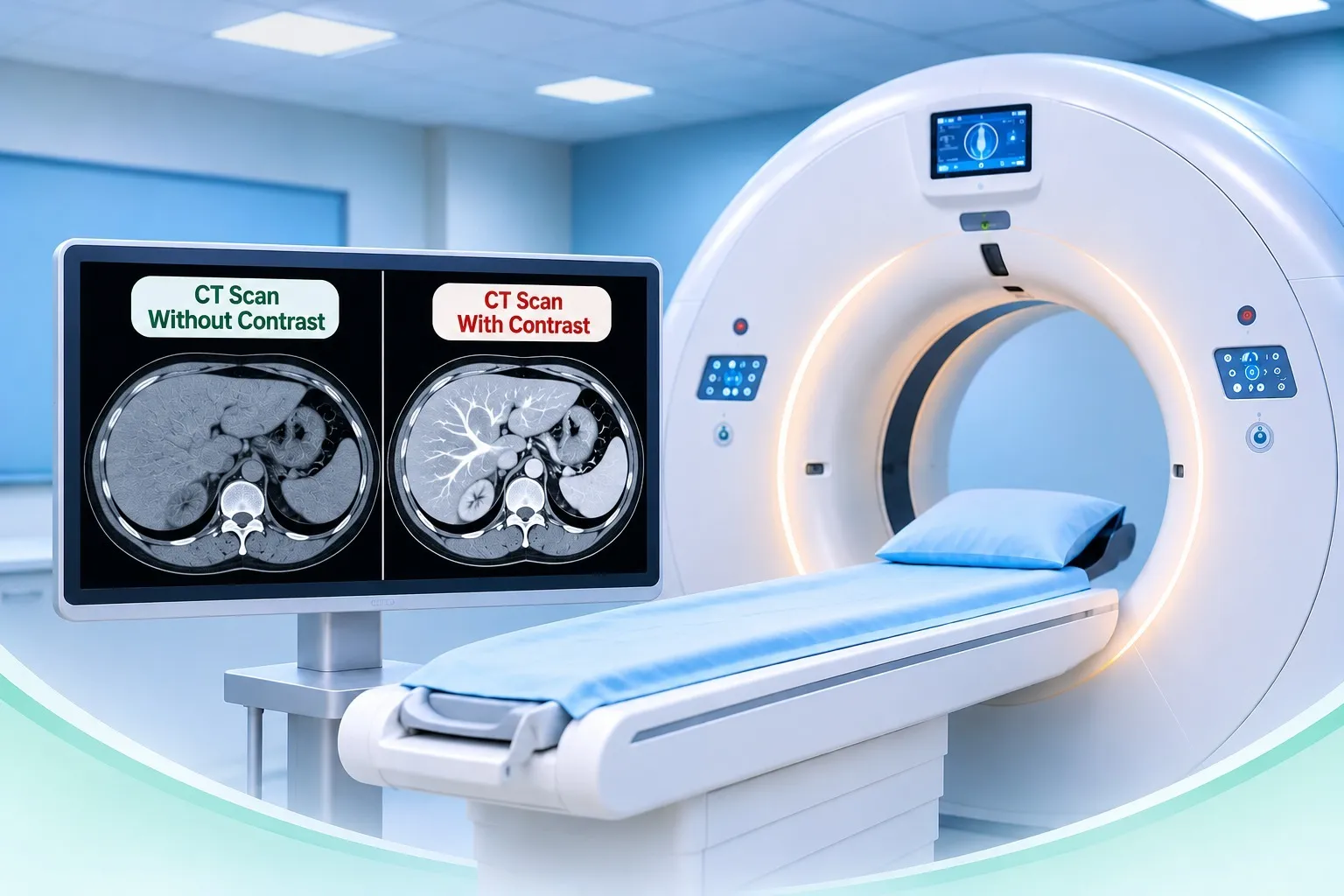
Why is contrast used? It highlights blood vessels, organs and abnormal tissue, making it easier to see tumours, infections, inflammation, clots and bleeding.
When is contrast necessary? It depends on the question your doctor is asking:
| Usually without contrast | Usually with contrast |
|---|---|
| Head injury, suspected brain bleed | Cancer detection and staging |
| Kidney stones | Abdominal infections and abscesses |
| Fractures and bone injuries | Suspected blood clot in the lungs |
| Lung nodule follow-up | Blood vessel problems (CT angiography) |
What precautions should you take? Tell your team about kidney disease, diabetes, previous contrast reactions, asthma or thyroid problems. Most reactions are mild, such as nausea, itching or a rash, and serious reactions are rare. Staff are trained to treat them immediately. Breastfeeding mothers can usually continue feeding normally; ask your doctor if you have concerns.
Is a CT Scan Safe?
Yes, when it is medically needed. For most patients, the benefit of an accurate, timely diagnosis far outweighs the small risk.
Radiation exposure: CT uses ionising radiation, measured in millisieverts (mSv). Everyone receives natural background radiation of about 3 mSv a year. Typical adult doses are roughly:
| Scan | Approximate dose | Similar to natural background radiation over |
|---|---|---|
| Chest X-ray | 0.1 mSv | 10 days |
| CT brain scan | 2 mSv | 8 months |
| Chest CT scan | 7 mSv | 2 years |
| CT abdomen and pelvis | 10 mSv | 3 years |
Source: RadiologyInfo.org (RSNA and ACR). Actual doses vary by scanner, protocol and body size.
Possible risks: a very small increase in lifetime cancer risk with high or repeated exposure, reactions to contrast dye, and rarely, kidney strain from contrast in people with existing kidney disease.
Is a CT scan safe during pregnancy? Always tell your doctor if you are or might be pregnant. Scans of the head, chest or limbs expose the baby to very little radiation. Scans of the abdomen and pelvis give more exposure, so doctors consider ultrasound or MRI first. In an emergency, a CT scan is done if it is the best way to protect the mother's health.
How is radiation minimised? Modern scanners adjust the dose to the patient's size, use advanced image reconstruction, and scan only the area needed. Children receive specially reduced-dose settings.
Why avoid unnecessary repeat scans? Radiation adds up over a lifetime. Always carry your previous scans and reports, and tell your doctor about recent imaging, so a repeat scan can be avoided where possible.
CT Scan vs X-Ray vs MRI
Each test has its strengths. Your doctor chooses the one most likely to answer the clinical question safely.
| Feature | X-Ray | CT Scan | MRI |
|---|---|---|---|
| How it works | A single X-ray beam makes a flat 2D image | Rotating X-rays and a computer make detailed cross-sectional slices | Strong magnet and radio waves make detailed images |
| Radiation | Very low | Low to moderate | None |
| Examination time | A few minutes | Usually under 5 minutes of scanning | 20 to 60 minutes |
| Best for | Fractures, chest infections, joint and dental problems | Emergencies, head injury, bleeding, lungs, abdomen, cancer staging | Brain, spinal cord, ligaments, cartilage and soft tissue detail |
| Common investigations | Broken bones, pneumonia, arthritis | Stroke, internal bleeding, appendicitis, kidney stones, lung nodules, pulmonary embolism | Slipped disc, tumours, multiple sclerosis, sports injuries |
| Points to note | Limited soft tissue detail | Contrast may be needed; avoid unnecessary repeats | Not suitable with some metal implants or pacemakers; noisy and enclosed |
When Will You Get Your CT Scan Report?
How radiologists review the images: After the scan, hundreds of images are sent to a radiologist, a doctor specialised in reading medical images. They review the scan slice by slice and in 3D, and compare it with your previous scans where available.
How the report is prepared: The report usually describes how the scan was done, the findings in each organ, and an "impression" that summarises the most important results and any suggested next steps.
Reporting time at Prayag Hospital: In emergency settings, CT reports are prioritised so that treatment is not delayed. For routine scans, our radiology team will tell you when your report will be ready.
Why discuss results with your doctor? A report can contain technical terms and incidental findings that may sound worrying but are often harmless. Your doctor will interpret the results alongside your symptoms, examination and other tests, and explain what they mean for your treatment.
CT Scan Services at Prayag Hospital
Prayag Hospital, a multi-speciality hospital at J-206/A-1, Sector 41, Noida (near Noida City Centre), offers diagnostic imaging services through its radiology department. Having CT scanning within a multi-speciality hospital means your scan, report and specialist consultation can happen under one roof, with emergency, orthopaedic, neurology, surgery and internal medicine teams on hand.
Booking an appointment: To enquire about a CT scan in Noida, appointment slots or preparation instructions, call Prayag Hospital at 0120 - 4021900 or visit the Radiology Department at Prayag Hospital. Please bring your doctor's prescription and previous reports when you come for your scan.
Conclusion
A CT scan is a fast, painless and highly detailed imaging test that helps doctors diagnose injuries, infections, bleeding, stones and cancers with confidence. A chest CT scan looks closely at the lungs and heart, a CT scan abdomen examines the digestive and urinary organs, and a CT brain scan is the first choice in head injury and suspected stroke.
With the right preparation, honest communication about allergies, kidney health and pregnancy, and a scan done only when it is needed, a CT scan is a safe and valuable step towards the right treatment. If your doctor has advised a CT scan, the radiology team at Prayag Hospital, Noida, can guide you through every step.